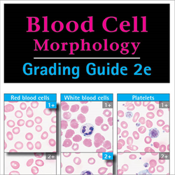
Blood Cell Morphology Grading Guide


Blood Cell Morphology Grading Guide By Gene Gulati
By Gene Gulati, 96 pp with illus, Chicago, Illinois, American Society for Clinical Pathology Press, 2009. Blood Cell Morphology: Grading Guide is a reference guide for grading red blood cell abnormalities, white blood cell abnormalities, and platelet morphology. The purpose of the book is to provide a practical approach that will make the evaluation of cell morphology in the manual differential white blood cell count and peripheral blood smear more systematic and consistent among laboratory professionals through the use of a grading system. This reference guide is divided into 4 sections. Section I discusses general considerations of grading blood cell morphology. Section II focuses on the specifics of grading individual red blood cell abnormalities. The grading of white blood cell abnormalities is described in Section III, and platelet morphology is discussed in Section IV.
In each section, grading is categorized from 1+ to 4+. Specific grading parameters are described for each abnormality and are illustrated by a photomicrograph taken at X1000 magnification, unless otherwise indicated, from a peripheral blood smear stained with Wright or Wright Giemsa. Section I: General Considerations Common hematologic tests performed in the clinical laboratory include the manual differential leukocyte count and morphologic verification of the automated complete blood cell count. Abnormal morphologic findings are reported in various ways, including with the use of such terms as present or absent, and semi-quantitatively, as slight, moderate, or marked. Although there is no evidence that either reporting system is superior to the others, the author emphasizes that maintaining consistency within a chosen system is good clinical practice and is also recommended by laboratory-accrediting agencies. To achieve consistency in grading, this book defines a grading system (1+ to 4+) for several abnormalities based on the relative degree of abnormality in individual cells, the relative fraction of cells with the abnormality, or (preferably) a combination of both.
Section II: Red Blood Cell Morphology Section II discusses the specifics of grading individual red blood cell abnormalities and defines a grading system described in words and images for anisocytosis, poikilocytosis, microcytosis, macrocytosis, hypochromia, polychromasia, blister cells, target cells, teardrop cells, schistocytes, sickle cells, spherocytes, acanthocytes, echinocytes, elliptocytes, stomatocytes, Howell-Jolly bodies, basophilic stippling, Pappenheimer bodies, rouleaux, and agglutination. For each red blood cell abnormality, the definition and associated clinical correlations are provided. In addition, correlations with automated results generated by the analyzer, such as red blood cell distribution width, mean corpuscular volume, and mean corpuscular hemoglobin concentration, are provided where applicable. For each abnormality, a grading system defined as occasional, 1+,2+,3+,or 4+ is provided, and parameters are set based on the abnormal cells as a percentage of all red blood cells for most cases, with the exception of anisocytosis, which is graded based on how large the representative largest red blood cell is compared with the representative smallest red blood cell. Additional grading criteria are given for some abnormalities, including microcytosis, which takes into account how the size of the representative smallest red blood cell compares to a normal red blood cell; macrocytosis, in which the size of the largest representative red blood cell is compared with a healthy red blood cell; and hypochromia, in which the size of the central pale area as a fraction of the diameter of the total area of the red blood cell is taken into consideration.
For each abnormality, the grading criteria are clearly defined and the corresponding color images are included to illustrate each abnormality according to grade. Section III: White Blood Cell Morphology Section III provides guidelines for grading toxic granulation, toxic vacuolation, Dohle inclusion bodies, hypersegmentation, hyposegmentation, agranular/hypogranular granulocytes, cytoplasmic fragments/ agranular or hypogranular platelets, and smudge cells. For each topic, clinical correlations associated with each morphologic finding are provided. Grading is classified as occasional, 1+, 2+, 3+, or 4+. For several abnormalities, grading is based on neutrophils plus bands with the specified abnormality as a percentage of all neutrophils and bands. Additional criteria are set for grading toxic granulation (which takes into account the average size and density of granules), toxic vacuolation (in which the average number of vacuoles per neutrophil and band are considered), and Dohle inclusion bodies (which takes into account the average number of Dohle inclusion bodies per neutrophil and band).
The grading of hypersegmentation is divided into 3 systems based on the average number of nuclear lobes, the percentage of neutrophils with 5 lobes, and the percentage of neutrophils with 6 lobes. The grade is determined for hyposegmentation, by the percentage of neutrophils with bilobed and unilobed nuclei; for hypogranularity, by the number of hypogranular cells as a percentage of all granulocytes; for cytoplasmic fragments and hypogranular platelets, as a percentage of the sum of all platelets and cytoplasmic fragments; and for smudge cells, based on smudge cells as a percentage of all white blood cells.
Blood Cell Morphology: Grading Guide serves as a comprehensive reference for grading red blood cell abnormalities, white blood cell abnormalities, and platelet morphology. This book provides clearly understandable parameters for the grading of several blood cell abnormalities and also serves as a review of blood cell morphology. Order # 6555 Author: Gene Gulati, PhD, SH(ASCP) New Second Edition Gulati’s updated, comprehensively illustrated guide makes the process of grading blood cell morphology more immediately practical for laboratory professionals - and more meaningful for patient management. Feb 24, 2014 - guidelines, and the advancements in hematology analyzers, the methods of reporting or grading abnormal red blood cell morphol- ogy still vary.
Color photomicrographs are provided for each entity to illustrate the grade. Section IV: Platelet Morphology Section IV provides guidelines for grading platelet morphology.

Blood Cell Morphology Grading Guide Pdf
This section provides a grading system for giant platelets, large platelets, agranular or hypogranular platelets and cytoplasmic fragments, and platelet satellitosis. Each morphologic category is graded as occasional, 1+, 2+, 3+, or 4+ and is categorized based on the abnormal platelets as a percentage of all platelets, with the exception of platelet satellitosis, which is graded based on neutrophils plus bands with platelet rosettes as a percentage of all neutrophils and bands or as the fraction of individual cell surfaces surrounded by platelets. Additional grading criteria for giant platelets are based on the size (diameter) of individual giant platelets as compared with healthy red blood cells. Color photomicrographs are provided for each platelet abnormality to illustrate the features of the grading system. Summary Blood Cell Morphology: Grading Guide serves as a comprehensive reference for grading red blood cell abnormalities, white blood cell abnormalities, and platelet morphology.
This book provides clearly understandable parameters for the grading of several blood cell abnormalities and also serves as a review of blood cell morphology. One of the greatest strengths of this book is the use of color photomicrographs to illustrate the features of each grade as it applies to the specific abnormal finding. The layout for each section is easy to follow. The grading systems are applicable to everyday practice, and the quality of the illustrations issuperb.Inaddition,theconstruction of the book, including the spiral binder and sleek design, allows for ease and comfort of use in any setting. This book is useful for laboratory professionals, including students, teachers, and practitioners. JESALYN JAHARA TAYLOR, MD Houston, Texas.